Slider

Science

SCITECH

AMAZING FACTS

NATURE SPACE

Psychology

Health Benefits of Sex

Regular sex cannot be underestimated as a factor for reducing stress, bolstering self-esteem and fostering feelings of intimacy and bonding between partners. But the real point of this article is the fact that a healthy sex life can provide for a longer, healthier and, most would agree, more enjoyable life. Among the many health benefits of sex are:


1. according to study, sex is the equivalent of playing doubles tennis or walking uphill.
2. sex burned 4 calories per minute for men and 3 per minute for woman, during sessions that ranged from 10 to 57 minutes, including foreplay. (the average was 25 minutes) man burned about 9 calories per minute jogging and woman about 7. sex is important for physical fitness
3.  sex increase your body's dopamine levels- the hormone that improve your memory and motivation.    
4. it helps you relax, lowering the stress levels in your body.lower blood pressure and overall stress reduction
5. when you have sex, the cuddle hormone oxytonic is released. The oxytonic released during orgasm also promotes sleep, according to research.
6. A sex life is also good for your heart. Besides being a great way to raise your heart rate, sex helps keep your estrogen and testosterone levels in balance.
“When either one of those is low you begin to get lots of problems, like osteoporosis and even heart disease,” Pinzone says.
Having sex more often may help. During one study, men who had sex at least twice a week were half as likely to die of heart disease as men who had sex rarely.
7. having sex once or twice a week has been linked with higher levels of an antibody called immunoglobulin A or lgA, which can protect you from getting colds and other infections.
8. sex help increase the release of the hormone testosterone , which in turn improves your energy level and strengthens vital body parts , like your muscles, bones and heart.
9.  frequent ejaculations , especially in 20-something men, may reduce the risk of prostate cancer later in life.
10. Sexual activity releases pain-reducing hormones and has been found to help reduce or block back and leg pain, as well as pain from menstrual cramps, arthritis and headaches. One study even found that sexual activity can lead to partial or complete relief of headache in some migraine and cluster-headache patients.

Gene Replacement and Transgenic Animals

Gene Replacement and Transgenic Animals

The goal of modern molecular cell biology is nothing short of understanding the biochemical, cellular, and organismal functions of all the proteins encoded in the . In the preceding sections, we have discussed the isolation and analysis of mutants, the genetic  of mutations, and finally the isolation and cloning of -defined genes. This approach can provide valuable information about the molecular mechanisms underlying the cellular processes affected by the original mutations and the functions of the normal proteins encoded by the affected genes. As discussed in Chapter 7, however, many genes have been identified based on the biochemical properties of their encoded , the sequence similarity of the encoded protein with proteins of known function, or their interesting patterns of  in . In the absence of mutant forms of such genes, their in vivo functions may be unclear. By mutating a specific   and then replacing the normal copy in the genome with a mutant form, scientists can assess its in vivo function. This technique, referred to as gene-targeted knockout, or simply “knockout,” is in essence the reverse of the approach described in the previous sections. The process of isolating normal genes to be mutated will be greatly simplified as sequencing of the genomes of several model organisms and of the human genome progresses (Chapter 7). Whether starting from a normal protein or sequenced genome, this approach can be summarized as follows:
Image ch8fu3.jpg
Other techniques permit the introduction of foreign genes or altered forms of an endogenous  into an organism. For the most part, these techniques do not result in replacement of the endogenous gene, but rather in the integration of additional copies of it. Such introduced genes are called transgenes; the organisms carrying them are referred to as . Transgenes can be used to study organismal function and  in a variety of different ways. For instance, genes that are normally expressed at specific times and places during development can be genetically engineered  to be expressed in different tissues at different times and then reintroduced into the animal to assess the cellular and organismal consequences. For example, theAntennapedia (Antp) gene in Drosophila normally controls leg development, but misexpression of this gene in the developing antenna transforms it into a leg.
The production of both -targeted knockout and  animals makes use of techniques for mutagenizing cloned genes  and then transferring them into eukaryotic cells. We briefly describe these procedures first, then discuss the production and uses of knockout and transgenic organisms.

Specific Sites in Cloned Genes Can Be Altered in Vitro

Specific sequences in cloned genes can be altered  and then introduced into experimental organisms. This approach has been exploited primarily to study two questions. First, what is the relationship between the structure of a particular  and its biological function? And second, what are the specific  sequences required to determine the  pattern of a particular?
A variety of enzymatic and chemical methods are available for producing site-specific mutations . In recent years, however, the most common methods use specific oligonucleotides as mutagens. Because oligonucleotides of any desired sequence can be chemically synthesized (see Figure 7-9), oligonucleotide-based mutagenesis can generate precisely designed deletions, insertions, and point mutations in a  sequence. Figure 8-29 illustrates the use of this strategy to produce a deletion.
Figure 8-29. In vitro mutagenesis of cloned genes with chemically synthesized oligonucleotides.

Figure 8-29

In vitro mutagenesis of cloned genes with chemically synthesized oligonucleotides. In this example, a cloned gene that normally contains three segments (A, B, and C) is mutagenized (more...)

DNA Is Transferred into Eukaryotic Cells in Various Ways

Production of both knockout and  organisms requires the transfer of  into eukaryotic cells. Many types of cells can take up DNA from the medium. Yeast cells, for instance, can be treated with enzymes to remove their thick outer walls; the resulting spheroplasts will take up DNA added to the medium. Plant cells also can be converted to spheroplasts, which will take up DNA from the medium. Cultured mammalian cells take up DNA directly, particularly if it is first converted to a fine precipitate by treatment with calcium ions. Another popular method for introducing DNA into yeast, plant, and animal cells is calledelectroporation. Cells subjected to a brief electric shock of several thousand volts become transiently permeable to DNA. Presumably the shock briefly opens holes in the cell  allowing the DNA to enter the cells before the holes reseal. DNA also can be injected directly into the nuclei of both cultured cells and developing embryos.
Once the foreign  is inside the host cell, enzymes that probably function normally in DNA repair and  join the fragments of foreign DNA with the host cell’s chromosomes. Since only a relatively small fraction of cells take up DNA, a selective technique must be available to identify the  cells. In most cases the exogenous DNA includes a  encoding a selectable marker such as drug resistance. The introduced DNA can insert into the host  in a highly variable fashion showing no site specificity, can replace an endogenous gene by homologous recombination, or can remain as an independent extrachromosomal DNA molecule referred to as an episome.

Normal Genes Can Be Replaced with Mutant Alleles in Yeast and Mice

Gene knockout is a technique for selectively inactivating a  by replacing it with a mutant  in an otherwise normal organism. This technique of disrupting gene function, which has been widely used in yeast and mice, is a powerful tool for unraveling the mechanisms by which basic cellular processes occur.

Gene Knockout in Yeast

After foreign, or exogenous, yeast  is taken up by  yeast cells,  generally occurs between the introduced DNA and the homologous chromosomal site in the recipient cell. Because of this specific, targeted recombination of identical stretches of DNA, called homologous recombination, any  in yeast chromosomes can be replaced with a mutant  (Figure 8-30). The resulting  yeast cells, carrying one mutant allele and one wild-type allele, generally grow normally. To determine whether the knocked-out gene controls an obligatory function, recombinants containing the mutant allele on one are treated to induce  and sporulation; each diploid cell produces four  spores, which are tested for viability. One of the first genes tested in this way was the one encoding , a prominent cytoskeletal  in yeast and higher organisms. Haploid yeast spores without a normal actin gene cannot grow (Figure 8-31).
Figure 8-30. Replacement of the normal HIS3 gene (blue) by homologous recombination with a mutant his3 gene (yellow) in the yeast S. cerevisiae..

Figure 8-30

Replacement of the normal HIS3 gene (blue) by homologous recombination with a mutant his3 gene (yellow) in the yeast S. cerevisiae.. Recombinant (more...)
Figure 8-31. Demonstration that actin gene is required for yeast viability by gene-targeted knockout.

Figure 8-31

Demonstration that actin gene is required for yeast viability by gene-targeted knockout. A recombinant plasmid containing the URA3 gene, a selectable marker, and a mutant actin gene (more...)
This technique also is useful in assessing the role of proteins identified solely on the basis of  sequence. For instance, the entire sequence of the S. cerevisiae  has been determined. As described in Chapter 7, analysis of genomic sequences can identify stretches of DNA that exhibit long open reading frames or  to genes encoding known proteins; such stretches are likely to be transcribed and translated into as yet unidentified proteins (see Figure 7-31). Gene knockouts can be used to determine whether such regions are important for specific cellular functions that are phenotypically detectable. This technique thus provides a powerful approach to identifying and studying new genes and the proteins that they encode.
This -knockout approach already has been used to analyze yeast  III. Analysis of the  sequence indicated that this chromosome contains 182 open reading frames of sufficient length to encode proteins longer than 100 amino acids, which is assumed in this analysis to be the minimum length of a naturally occurring . The sequences of the proteins that could be encoded by 116 of these putative protein-coding regions exhibited no obvious  to any known proteins. Gene knockout of 55 of these regions showed that 3 were required for viability; further analysis of 42 nonessential genes revealed that 14 showed a mutant  and 28 did not. The large number of putative genes with no detectable mutant phenotype is quite surprising. In some cases the lack of a phenotype could indicate the existence of backup or compensatory pathways in the cell. Alternatively, the mutations may give rise to subtle defects that would require more in-depth phenotypic analysis to uncover.

Gene Knockout in Mice

Gene-targeted knockout mice are a powerful experimental system for studying , behavior, and physiology; they also may be useful model systems for studying certain human genetic diseases. The procedure for producing -targeted knockout mice involves the following steps:
1.
Mutant alleles are introduced by homologous  into embryonic stem (ES) cells.
2.
ES cells containing a knockout  in one  of the  being studied are introduced into early mouse embryos. The resultant mice will be  containing tissues derived from both the transplanted ES cells and the host cells. These cells can contribute to both the germ-cell and somatic-cell populations.
3.
Chimeric mice are mated to assess whether the  is incorporated into the .
4.
Mice each  for the knockout  are mated to produce  knockout mice.
The isolation and culture of embryonic stem cells, which are derived from the blastocyst, are illustrated in Figure 8-32. These cells can be grown in culture through many generations. Exogenous  containing a mutant  of the  being studied is introduced into ES cells by . The introduced DNA recombines with chromosomal sequences in about 1 cell out of 100 (i.e., 1 percent  frequency). In some cells, the added DNA recombines with the homologous chromosomal site, but recombination at other chromosomal sites (i.e., nonhomologous recombination) occurs 103 – 104 times more frequently. The small fraction of cells in which homologous recombination takes place can be identified by a combination of positive and negative selection: positive selection to identify cells in which any recombination occurs and negative selection to remove cells in which recombination takes place at nonhomologous sites.
Figure 8-32. Preparation of embryonic stem (ES) cells.

Figure 8-32

Preparation of embryonic stem (ES) cells. Fertilized mouse eggs divide slowly at first; after 41/2 days, they form the blastocyst, a hollow structure composed of about 100 cells surrounding (more...)
For this selection scheme to work, the  constructs introduced into ES cells need to include, in addition to sequences used to knock out the  of interest, two selectable marker genes (Figure 8-33). One of these additional genes (neor) confers neomycin resistance; it permits positive selection of cells in which either homologous (specific) or nonhomologous (random) has occurred. The second selective gene, the thymidine  gene from herpes simplex  (tkHSV) confers sensitivity to ganciclovir, a cytotoxic  analog; this gene permits negative selection of ES cells in which nonhomologous recombination has occurred. Only ES cells that undergo homologous recombination (i.e., gene-targeted specific insertion of the DNA construct) can survive this selection scheme.
Figure 8-33. Isolation of mouse ES cells with a gene-targeted disruption by positive and negative selection.

Figure 8-33

Isolation of mouse ES cells with a gene-targeted disruption by positive and negative selection. (a) When exogenous DNA is introduced into ES cells, random insertion via nonhomologous (more...)
Once ES cells  for a knockout  in the  of interest are obtained, they are injected into a recipient mouse blastocyst, which subsequently is transferred into a surrogate pseudopregnant mouse (Figure 8-34). If the ES cells also are for a visible marker trait (e.g., coat color), then chimeric progeny carrying the knockout mutation can be identified easily. These are then mated with mice homozygous for another  of the marker trait to determine if the knockout mutation is incorporated into the . Finally, mating mice, each heterozygous for the knockout allele, will produce progeny homozygous for the knockout mutation.
Figure 8-34. General procedure for producing gene-targeted knockout mice.

Figure 8-34

General procedure for producing gene-targeted knockout mice. Embryonic stem (ES) cells heterozygous for a knockout mutation in a gene of interest (X) and homozygous for a (more...)

Cell-Type-Specific Gene Knockout in Mice

In most cases, investigators are interested in examining the effects of knockout mutations in a particular region of the mouse, at a specific stage in , or both. Since most genes function in different parts of the organism and at different times, a knockout mouse may die or have defects in various tissues prior to the stage to be analyzed. To address this problem, mouse geneticists have devised a clever technique using site-specific   sites (called loxP sites) and the , calledCre, that catalyzes recombination between them. The loxP-Cre recombination system is present in bacteriophage P1, but also promotes recombination when placed in mouse cells.
Homologous  strategies discussed in the previous section are used to obtain mice in which loxP sites are inserted so they flank the  of interest or an essential . Since the inserted loxP sites are not within exons, they do not by themselves disrupt gene function. Transgenic mice also are prepared carrying the cre gene linked to a cell-type-specific . As depicted in Figure 8-35, mating of these two types of mice will yield progeny that carry the gene of interest modified by insertion of flanking lox P sites and the cre gene controlled by a cell-type-specific promoter. In these mice, recombination between the loxP sites, which disrupts the gene of interest, will occur only in those cells in which the promoter is active and therefore producing the Cre necessary to induce the recombination.
Figure 8-35. Cell-type-specific gene knockouts using the loxP-Cre recombination system.

Figure 8-35

Cell-type-specific gene knockouts using the loxP-Cre recombination system. Two loxP sites are inserted on each side of an essential exon (2) of the gene of interest (i.e., gene X) (more...)
One important example of this technique comes from studies on learning and memory. Earlier pharmacological and physiological studies had indicated that normal learning requires a specific  , the NMDA class of glutamate receptors, in a specific region of the brain called the hippocampus. But mice in which the  encoding an NMDA receptor subunit was knocked out died neonatally, precluding analysis of the receptor’s role in learning. Cell-type-specific inactivation of the receptor was achieved by constructing mice carrying a Cre gene expressed in a subclass of hippocampal neurons and two different alleles of the receptor subunit gene, an allelle containing the loxP sites and a conventional knockout . These mice survived to adulthood and showed learning and memory defects, confirming a role for these receptors in normal learning and memory.

Use of Knockout Mice to Study Human Genetic Diseases

Image med.jpgGene knockout can produce model systems for studying inherited human diseases. Such model systems are powerful tools for investigating the nature of genetic diseases and the efficacy of different types of treatment, and for developing effective  therapies to cure these often devastating diseases.
Recent studies on cystic fibrosis illustrate this use of the knockout technique. Cystic fibrosis, which afflicts about 1 in 2000 Caucasians, is caused by an autosomal   in the CFTR  (see Figure 8-17b). This gene was cloned by strategies, and the biochemical function of its encoded  studied. Using the human gene, researchers isolated the homologous mouse gene and subsequently introduced mutations in it. The gene-knockout technique was then used to produce  mutant mice, which showed symptoms (i.e., a ) similar to those of humans with cystic fibrosis. These knockout mice are currently being used as a model system for studying this genetic disease and developing effective therapies.

Foreign Genes Can Be Introduced into Plants and Animals

In the previous section we discussed techniques for replacing one form of a  with another through homologous . In this section we discuss methods for producing  organisms, which carry cloned genes that have integrated randomly into the host .
Transgenic technology has numerous experimental applications and potential agricultural and therapeutic value. For instance, dominantly acting alleles of -causing genes can be used to produce  mice, thus providing an animal model for studying cancer. In Drosophila, transgenes often are used to determine whether a cloned segment of  corresponds to a defined by . If the cloned DNA is indeed the gene in question, then introducing it as a  into a mutant fly will transform the mutant into a phenotypically normal individual. Transgenic plants may be commercially valuable in agriculture. Plant scientists, for example, have developed transgenic tomatoes that exhibit reduced production of ethylene, which promotes fruit ripening. The ripening process is delayed in these transgenic tomatoes, thus prolonging their shelf life. Finally, transgenic technology is a critical component in the burgeoning field of gene therapy for human genetic diseases.

Transgenic Mice

As noted in the discussion of knockout mice, specific integration of exogenous  into the  of mouse cells by homologous  occurs at a very low frequency. In contrast, the frequency of random integration of exogenous DNA into the mouse genome at nonhomologous sites is very high. Because of this phenomenon, the production of  mice is a highly efficient and straightforward process.
As outlined in Figure 8-36, foreign  containing a  of interest is injected into one of the two pronuclei (the male and female nuclei contributed by the parents) of a fertilized mouse egg before they fuse. The injected DNA has a good likelihood of being randomly integrated into the chromosomes of the  . Injected eggs then are transferred to foster mothers in which normal cell growth and  occurs. About 10 – 30 percent of the progeny will contain the foreign DNA in equal amounts (up to 100 copies per cell) in all tissues, including germ cells. Immediate breeding and backcrossing (parent-offspring mating) of the 10 – 20 percent of these mice that breed normally can produce pure  strains  for the .
Figure 8-36. General procedure for producing transgenic mice.

Figure 8-36

General procedure for producing transgenic mice. [See R. L. Brinster et al., 1981, Cell27:223.] View Movie: Creating a Transgenic Mouse
Image med.jpgNumerous examples of the use of  mice for studying various aspects of normal mammalian biology are presented in other chapters. They also provide a model system for studying disease processes. For example, many forms of cancer are promoted by normal cellular genes acting in a  fashion owing to their misregulated activity. Although transgenic mice carrying one of these genes, called myc, develop normally, tumors form at a high frequency. The observation that only a small number of cells expressing the  develop tumors supports a model in which additional genetic changes are necessary for tumors to form. These mice may provide an important tool for identifying those changes.

Transgenic Fruit Flies

Foreign  can be incorporated into the Drosophila germ-line  by the technique of P-element  (Figure 8-37). This technique makes use of a segment of the P element, a highly , which can transpose (jump) from an extrachromosomal element into a . (Mobile DNA elements are discussed in detail in Chapter 9.) Generally, this procedure results in incorporation of a single copy of the  into the Drosophila genome. In contrast,  mice carry multiple copies of the transgene incorporated into their chromosomes. In both organisms, however, the chromosomal insertion site is highly variable.
Figure 8-37. Generation of transgenic fruit flies by P-element transformation.

Figure 8-37

Generation of transgenic fruit flies by P-element transformation. The P element, a mobile genetic element, can move from one place in the genome to another. This movement (transposition) (more...)
Flies that develop from injected embryos will carry some germ cells that have incorporated the ; some of their progeny will carry the transgene in all somatic and germ-line cells, giving rise to pure  lines. Individuals carrying the transgene are recognized by  of a marker  (e.g., one affecting eye color) that is also present on the donor . Although the transgenes in Drosophila and mice insert in chromosomal sites different from the position of the corresponding endogenous gene, they usually are expressed in the right tissue and at the right time during . Examples of the importance of this technology for studying development are discussed in Chapter 14.

Transgenic Plants

Image plant.jpgIn nature, plant cells often live in close association with certain bacteria, which may provide a convenient vehicle for introducing cloned  into plants. Agrobacterium tumefaciens, for example, attaches to the cells of dicotyledonous plants and causes the formation of plant tumors known as galls. (Plants with two leaflets from each seed are calleddicotyledons, or dicots; plants with one leaflet are called monocots .) This bacterium introduces a circular DNA molecule, called theTi (-inducing) , into the plant cell in a manner similar to bacterial conjugation. The plasmid DNA then recombines with the plant DNA. Since the Ti plasmid has been isolated, new genes can be inserted into it using  techniques and the Ti genes causing tumors can be disrupted. The resulting recombinant plasmid can then transfer desired genes into plant cells (Figure 8-38).
Figure 8-38. Production of transgenic plants with recombinant Ti plasmids.

Figure 8-38

Production of transgenic plants with recombinant Ti plasmids. In nature, the Ti (tumor-inducing) plasmid in A. tumefaciens gains entry into a plant and is (more...)
An especially useful characteristic of plants for  studies is the ability of cultured plant cells to give rise to mature plants. Meristematic (growing) cells from dissected plant tissue or cells within excised parts of a plant will grow in culture to form callus tissue, an undifferentiated lump of cells. Under the influence of plant growth hormones, different plant parts (roots, stems, and leaves) develop from the callus and eventually grow into whole, fertile plants. When an agrobacterium containing a recombinant Ti infects a cultured plant cell, the newly incorporated foreign  is carried into the plant .
As noted above A. tumefaciens readily infects dicots (petunia, tobacco, carrot) but not monocots; reliable techniques for introducing genes into monocots are still being developed. Direct introduction of  by electroporation has been successful in rice plants, which are monocots, and the future looks bright for the manipulation of other commercially important monocotyledonous crop plants. Also available for -transfer experiments are cells of a tiny, rapidly growing member of the mustard family calledArabidopsis thaliana. This plant is well-suited to genetic analysis of a variety of developmental and physiological processes. It takes up little space, is easy to grow, and has a small , and genes defined by mutations can be cloned by  strategies.

SUMMARY

  •  Genes can be modified  by a variety of enzymatic and chemical methods. Modified genes can be incorporated into the  at their original genomic location by homologous , producing knockouts, or at different sites by nonhomologous recombination, producing transgenics.
  •  Any cloned yeast  or mouse gene can be mutated and used to generate gene-targeted knockouts in these organisms (see Figures 8-27 and 8-31). Use of the loxP-Cre  system from bacteriophage P1 permits production of mice in which a gene is knocked out in a specific tissue (see Figure 8-35).
  •  Clues about the normal function of a  can be deduced from the observed effects of disrupting it by the knockout technique. Mouse knockouts can provide models for human genetic diseases such as cystic fibrosis.
  •  Transgenic single-cell organisms, plants, and animals can be produced readily by several different methods. These modified organisms contain one or more copies of a cloned  integrated into in the .
  •  The  technique can be used to introduce a normal copy of a  into a mutant organism, thereby identifying a cloned  corresponding to a -defined gene. It also is used to study sequences necessary for , to develop mouse models of  forms of human diseases, to modify plants, and to investigate the relationship between the structure of a  encoded by a gene and its function.

Dogs Help Cheetahs Overcome Breeding Fears In New Zoo Project

LOS ANGELES — It may sound like a West Side Story-style love story, but some dogs love big cats.
Cheetahs are the fastest mammals in the world, but they also are the world's biggest scaredy-cats — so much so that they don't breed easily and are in danger of going extinct.
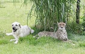
Some zoos are introducing dogs to calm the skittish cats and bring attention to their plight. They're pairing "companion dogs" with some cheetahs to serve as playmates and to provide the cats with guidance.

A Cheetah and Dog who are best friends

"It's a love story of one species helping another species survive," said Jack Grisham, vice president of animal collections at the St. Louis Zoo and species survival plan coordinator for cheetahs in North America.
Or, to quote Stephen Stills, it's a matter of loving the one you're with, he said.
"It is all about comforting and reassuring the cheetah," said Janet Rose-Hinostroza, animal training supervisor at the San Diego Zoo Safari Park — the top U.S. breeder of cheetahs in captivity. In the past 40 years, 135 cheetahs have been born at the park's breeding facility.
The cheetahs most often found at zoos and wildlife parks are not considered good breeding candidates, they don't relate well to other cheetahs, or they are abandoned by their mothers, Rose-Hinostroza said. But they seem to take easily to companion dogs and look to the dogs for play and example.
Of the 19 cheetahs at Safari Park, four have dogs. Four of the zoo's cheetahs also have dogs.
The dogs, usually from animal shelters, and cheetah pups generally are introduced when they are about 3 months old.
"In this relationship, the dog is dominant, but we look for dogs that want to be a buddy," Rose-Hinostroza said. "The dog always has the cat's back, but it's never the other way around. Dogs worry about their cats. They protect their cats."
One of the most popular draws at Safari Park is the 100-meter cheetah run where the public gets to see firsthand the speed of "nature's perfect sprinter."
"Speed is incredibly important. It is their survival technique, in a nutshell," Rose-Hinostroza said. "If they can't run, they won't survive. They are not equipped to be confrontational."
A cheetah's claws don't retract, so they have footing that takes them from "zero to 60 in 3.4 seconds," she said.
"That's faster than every single car on the market, and it only takes three steps," Rose-Hinostroza said.
Cheetahs use their tails like a rotor to balance while they are running. Their top speed is 60 to 70 mph, based on size, but they can run that fast only for 20 or 30 seconds. Extending that to a minute or more puts the animal in serious jeopardy of death.
"Overexertion, heat exhaustion can literally cook their organs at that speed," Rose-Hinostroza said. She added the average cheetah chase in the wild is 200 to 300 meters.
Safari Park's cheetahs chase a lure for 100 meters, a sprint that seldom exceeds 6 seconds.
A century ago there were 100,000 cheetahs in the wild, Grisham said. Today there are fewer than 12,000. The species has become extinct in at least 13 countries. There are about 280 captive cheetahs in zoos across the United States.
As captive efforts to save the species continue, Grisham worries there is no wild to send them home to because habitat is being swallowed up by developers and poachers are killing the cats for their fur.
Cheetahs live 12 to 15 years in captivity. Males weigh 120 to 150 pounds, and females 100 to 120 pounds.
The dogs come in all sizes. At Safari Park, the smallest and sweetest is Hopper, a male mutt who weighs 40 pounds. He's teamed with Amara, the toughest female cheetah on the team, Rose-Hinostroza said.
Cheetah females don't go into heat like other cats. Instead, they have to be brought into estrus by a male cheetah, the experts explained. That's why breeding is so hard — because they aren't social animals, they live independently, and they seldom hang out with one another.
Although the dogs and cats live together, they are not always with one another. Dogs have play dates with other dogs and humans. Mealtimes always are spent apart. The dogs eat kibble, and the cheetahs eat steak.
"The dogs are the bosses in these relationships," Rose-Hinostroza said. "If they ate together there would be one really fat dog and a really skinny cheetah."
One of Safari Park's dogs — the only non-shelter dog — is Yeti, an Anatolian shepherd. She works with two cheetahs — Johari and her brother Shiley.
No one is sure when the idea of cheetah dogs started, but Anatolian shepherds helped advance it. The San Diego Zoo was given a pair of cheetahs in 1981 on the condition they be given dogs because they were used to them.
A few decades ago, Dr. Laurie Marker, founder and executive director of the Cheetah Conservation Fund in the southern African nation of Namibia, brought Anatolian shepherds from Turkey and raised them to protect area goat herds.
"The Anatolian shepherd weighs up to 150 pounds and isn't afraid of anything," Grisham said. "They'll square off against lions and leopards. They don't always win, but they are very protective.
"Marker gave the dogs to farmers to protect their herds," Grisham said. When cheetahs came looking for dinner, the dogs scared the cats away and saved the farmer's goats. At the same time, the dogs saved the cats from being killed by the farmers. There was plenty of other food in the wild for the cats, including gazelles, impalas, springhares, birds, warthogs, kudu and hartebeest.
The dogs have helped cheetah conservation in Africa. "For the first time in 30 years, the cheetah population in the wild is on the rise because ranchers don't have to shoot them anymore. They don't need to shoot them. The dog is that effective at keeping the cheetah away from the herd," Rose-Hinostroza said.
Not every zoo that breeds cheetahs uses dogs. The St. Louis Zoo, where Grisham is based, has seven cheetahs but does not use dogs. More than 30 cubs have been born at that zoo.
Visitors can still watch the skittish cats and learn what all the fuss is about, Grisham said.
"It helps us understand the plight of animals in nature. In Africa, cheetahs were treated as vermin for years, like people in the United States treat coyotes," Grisham said.
Reference:

Top